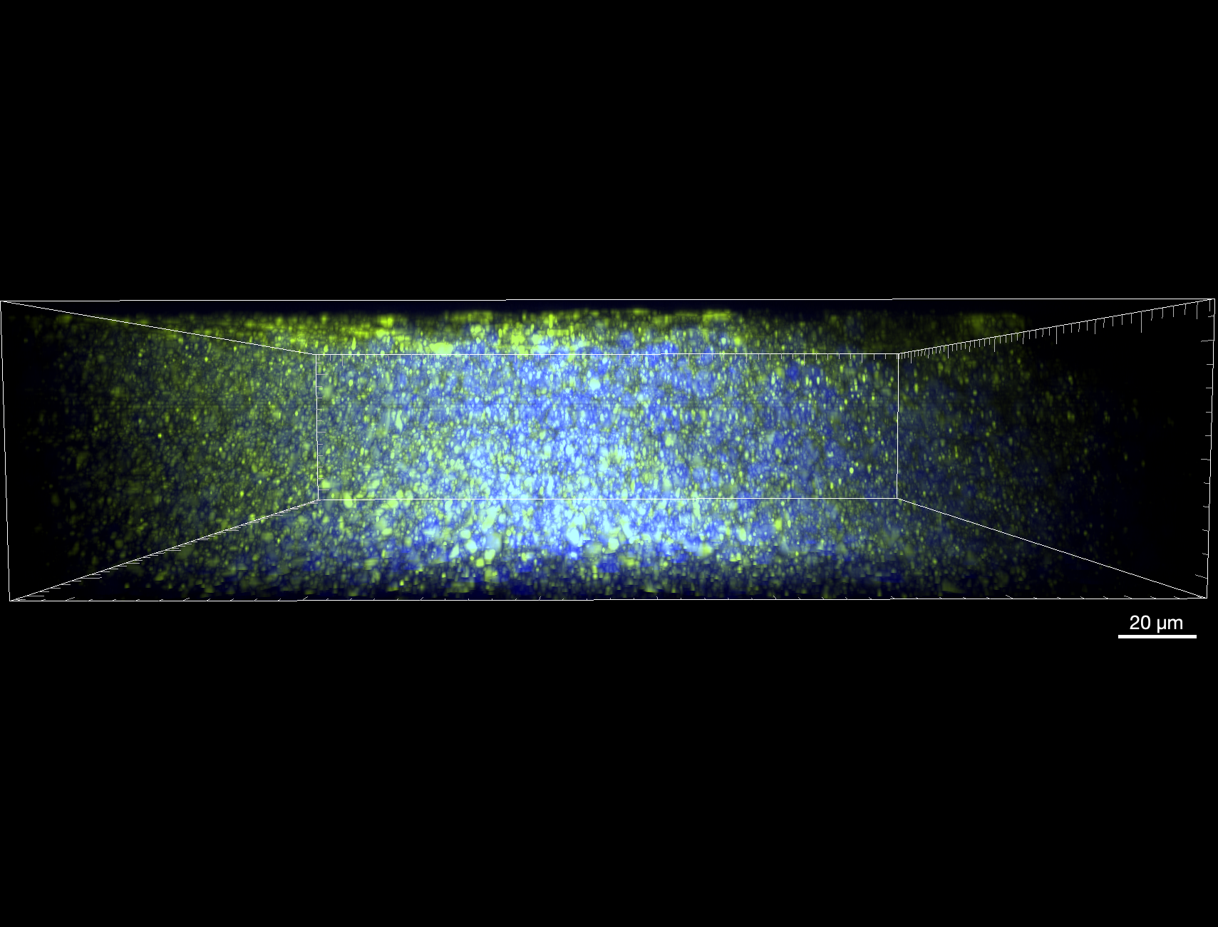
How does DNA microscopy reveal spatial biology in 3D?
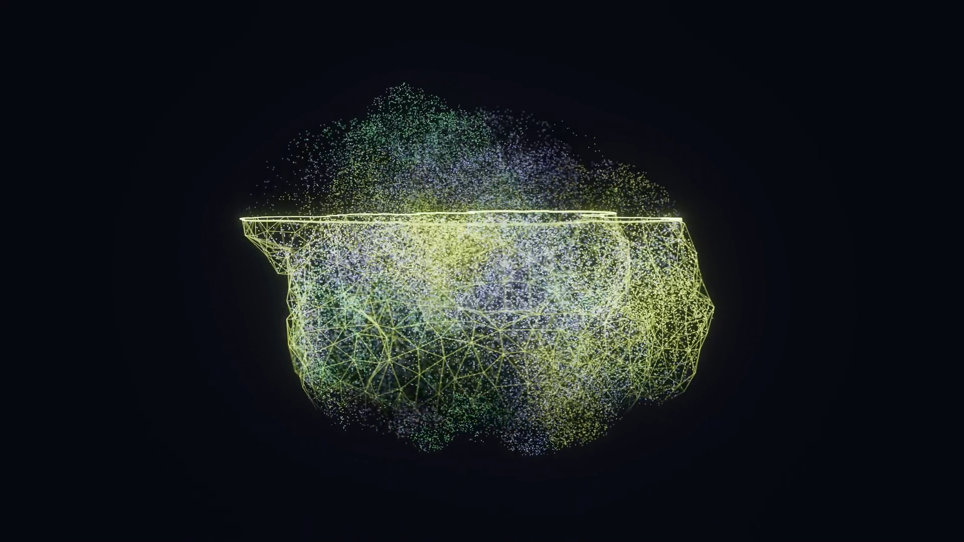
Traditional microscopy relies on light, lenses, and physical line-of-sight to reveal biological structures. But when studying complex 3D tissue architectures such as tumor microenvironments or intact developing organisms, optical limitations often force researchers to choose between slicing sample tissue into thin 2D sections or sacrificing molecular depth.
A landmark study published in Nature Biotechnology by Qian & Weinstein in 2025 (see the publication section) presents a new technological approach to overcome these limitations called volumetric DNA microscopy.
Rather than illuminating a sample from the outside, this technique uses a distributed network of DNA molecules to construct 3D spatial transcriptomic maps from the inside out.
An "Optics-Free" workflow
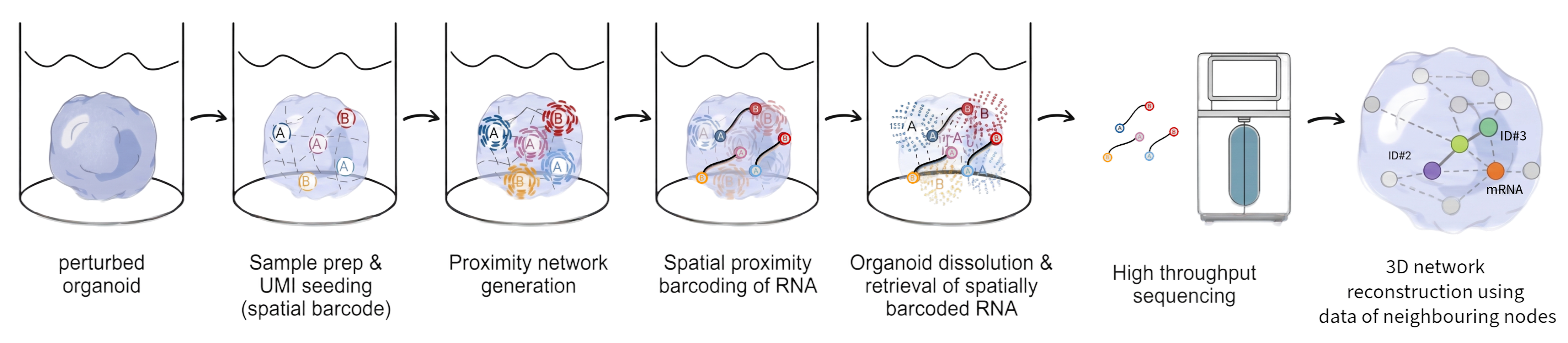
DNA microscopy shifts the focus from physical optics to molecular chemistry and high-throughput sequencing. The workflow being developed at Cubase Bio is being built from the foundational research published by Qian and Weinstein in Nature Biotechnology (2025) and we can distill it down to 4 main stages:
- Target RNA in Place: Inside an intact 3D cell culture or tissue sample, target RNA molecules remain fixed in their original physical positions.
- Molecular Tagging: Reverse transcription converts each target RNA into complementary DNA (cDNA) inside the sample. Each cDNA molecule is tagged with a Unique Molecular Identifier (UMI), which acts as a distinct spatial barcode.
- Proximity-Based reaction: The barcoded molecules generate copies creating a dense, localized cloud of identical barcode replicas anchored around the original target molecule. As neighboring barcode molecule clouds expand, adjacent clouds overlap where target molecules sit close together in physical space. The overlapping barcode strands form a Unique Event Identifier (UEI), creating a physical record confirming that two specific molecules were neighbours.
- Computational reconstruction: The tissue sample is dissolved, and all linked UMI–UEI DNA strands are retrieved and read on any standard Next-Generation DNA Sequencer. Our specialized software then mathematically decodes the sequencing data, reconstructing the 3D coordinates of every gene transcript in the sample.
Why true 3D spatial biology matters?
While traditional spatial transcriptomics relies heavily on 2D tissue sections, physical cuts can obscure the biological reality of complex structures in several critical ways:
Tumor-Immune interactions: a 2D slice can show immune cells near a tumor, but whether they're actually infiltrating, walled off by stroma, or excluded entirely often only resolves in 3D. A single section can make an immunologically "cold" tumor look "hot" (or vice versa) purely by where the cut happened to land.
Vascular and neural networks: Vessels and axons branch, loop, and travel out of plane constantly. 2D spatial can show a vessel appearing to end abruptly (interpreted as pruning or occlusion) when it simply exited the section. The same goes for tracing neural connectivity claims from serial 2D sections that can easily invert cause and effect.
Tissue architecture and invasion fronts: Whether a tumor is growing in cohesive sheets or invasive single-cell streams, or whether a duct or gland structure is intact versus breached, depends on the connectivity of the whole structure, something a single plane can badly misrepresent.
Sparse events and rare phenotypes: Rare cell types, micrometastases, or small clusters are easy to miss entirely if the 2D plane doesn't intersect them — a false negative rather than a misinterpretation, arguably the more dangerous failure mode, since nothing looks wrong on the slide.
Looking ahead with 3D spatial biology
As spatial biology shifts from 2D sections to intact 3D omic technologies, tools like volumetric DNA microscopy underscore a broader trend: leveraging sequencing technologies and computational models to solve physical measurement bottlenecks. This new workflow for implementing an optics-free approach to 3D spatial transcriptomics highlights the foundational technology we are developing to ensure this capability can be utilised in labs worldwide.