Seeing Inside Intact Organoids using a DNA Sequencer
Nearly 90% of drug candidates fail during clinical trials, largely because results from 2D cell cultures or animal models fail to translate to human biology. Animal models are not only costly and ethically challenging, but they frequently fail to capture human-specific cell signaling, tissue architecture, and disease mechanisms.
Patient-derived organoids have emerged as a powerful alternative for drug discovery and disease modelling under global regulatory pushes (such as the FDA Modernization Act) to reduce animal testing in the pre-clinical stages of drug development. However, adoption has been limited by a lack of scalable assay tools to probe organoids at high throughput. Animal testing can only be meaningfully reduced if scientists can extract deep, reproducible, high-throughput data from complex 3D organoid models.
The trade-off holding back 3D organoid analysis
3D organoid models offer human physiological relevance for drug discovery and disease modeling, yet traditional analysis methods fail to keep pace. Current workflows present a frustrating trade-off:
High-content 3D microscope imaging for spatial analysis requires 3–4 days per 384-well plate, generates 5–10 TB of data, and tracks only 3–4 markers.
Standard 2D sectioning requires researchers to pick, transfer, or painstakingly align individual organoids onto sample holders or chucks: a cumbersome process that threatens sample integrity and limits high-throughput screening.
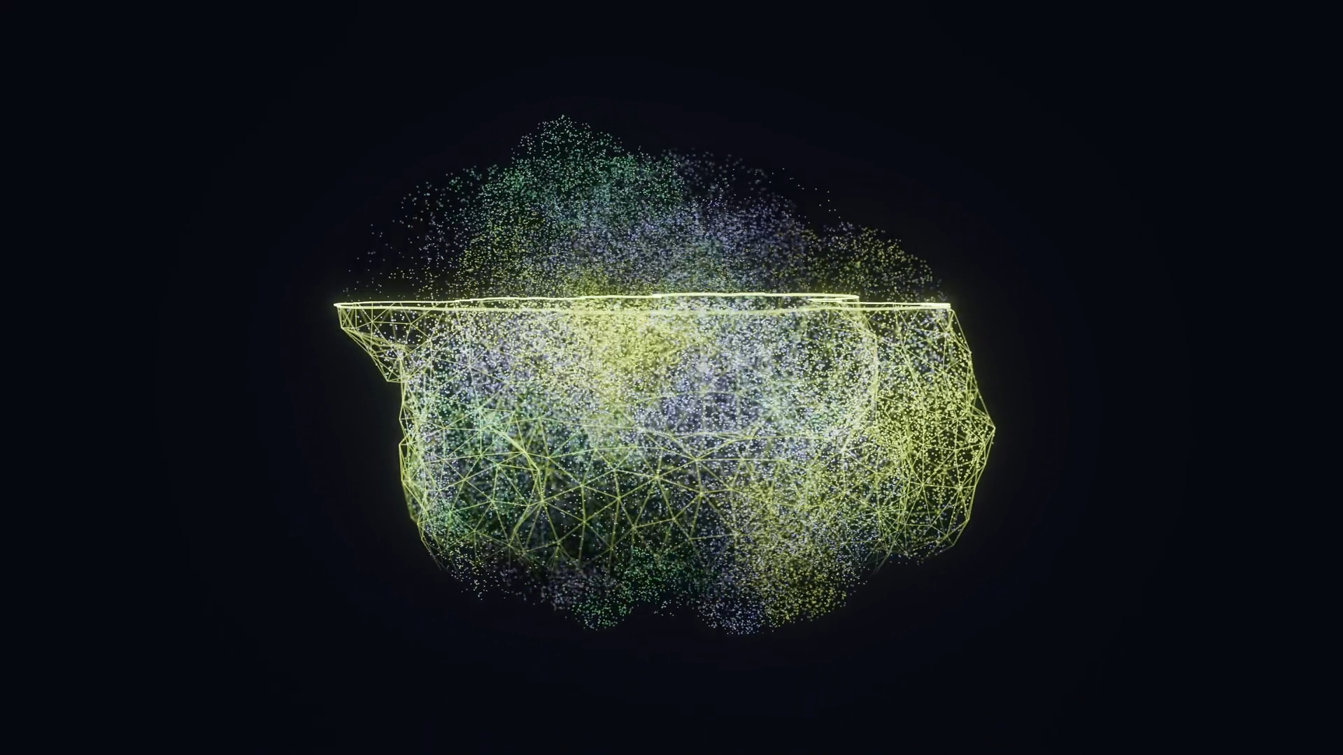
To solve this, we turned to DNA microscopy, a technique that replaces optical lenses with molecular reactions by combining sample preparation directly inside 384-well plates with a high-throughput DNA sequencing readout. By replacing heavy image files with compact X, Y, Z spatial coordinate data across all 384 wells, Cubase Bio enables 3D reconstructions of intact organoids across an entire plate.
Putting the technology to the test on human brain organoids
Beyond the zebrafish whole-organism dataset published in Nature Biotechnology by Qian and Weinstein (2025), Cubase Bio has generated proof-of-concept data applying the platform to a therapeutically relevant human 3D culture model: intact human brain organoids. This work is a validation study, demonstrating that the platform's core chemistry extends to an organoid workflow widely used in drug discovery and development.
Confirming reagent penetration into intact organoids
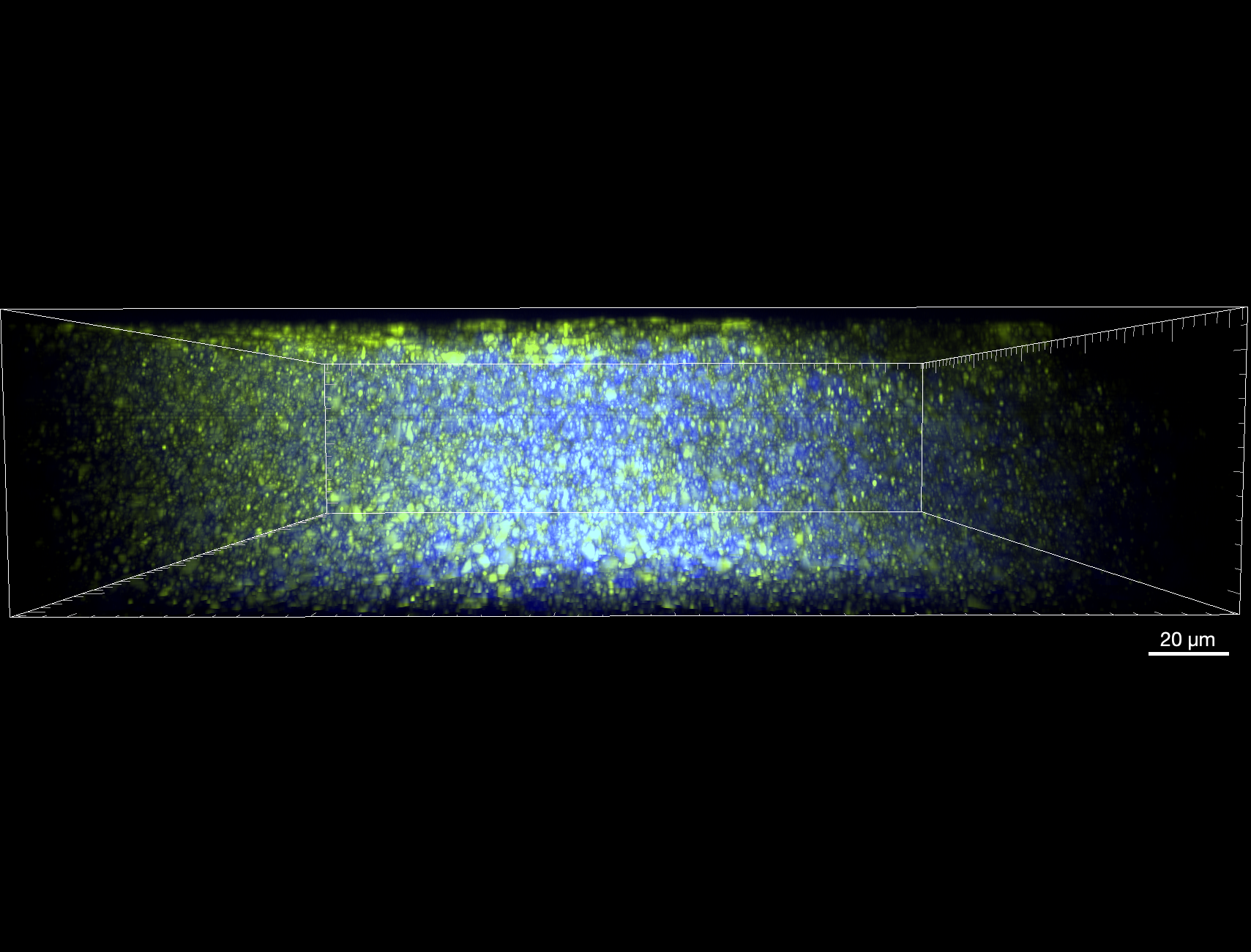
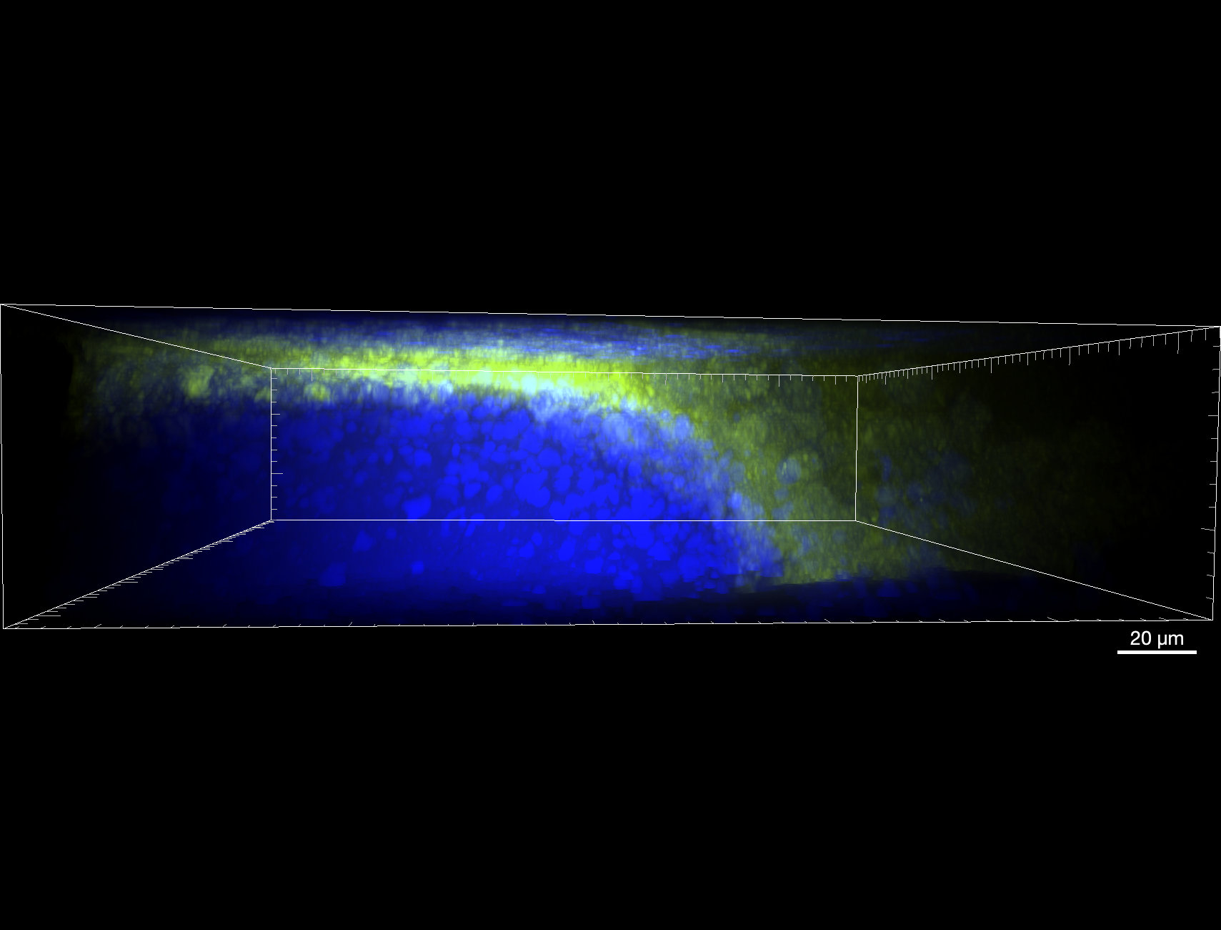
A central engineering challenge in moving from thin tissue sections to intact 3D samples is ensuring that the barcoding chemistry penetrates uniformly throughout the sample rather than remaining confined to its surface. Cubase Bio validated this directly using confocal microscopy on 1 mm³ human brain organoids obtained from BrainZell (Stockholm, Sweden). Initial preparations showed spatial array signal concentrated in a thin shell at the organoid periphery, with minimal signal reaching the organoid core (~400 µm deep, imaged at 60x). Following optimization of the sample preparation chemistry, spatial array signal co-localizes with nuclear staining throughout the full depth and volume of the organoid, confirming that the chemistry adequately labels the intact 3D structure rather than only its outer layers.
Mapping transcripts with a neurodevelopmental gene panel
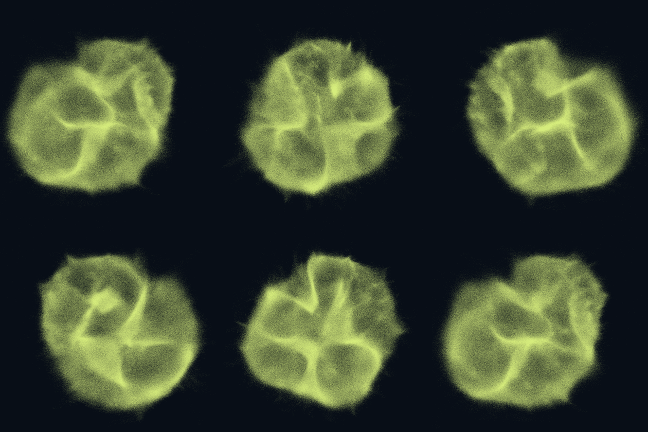
Using a targeted 50-gene panel focused on canonical neurodevelopmental markers — including VIM, VCAN, NES, MKI67, and DCX, spanning neural stem/progenitor identity, proliferation, and early neuronal differentiation — the platform generated 3.1 million barcoded molecular nodes and mapped 277,082 individual transcripts across the organoid. Representative z-slices through the organoid show spatially resolved expression of these markers, consistent with the known organization of proliferative and differentiating zones in developing neural tissue.
What's next: scaling to drug discovery and beyond
Because this workflow relies on chemical reactions and standard NGS readouts rather than complex microscopy hardware, it is uniquely suited to scale. Our vision for this platform includes:
High-Throughput Screening: Performing 3D spatial profiling directly in 96- and 384-well plates for drug candidate evaluation and New Approach Methodologies (NAMs).
Complex Models: Expanding beyond organoids and zebrafish to thick tissue samples, tumor core biopsies, and intact mouse organs.
By eliminating the need for physical sectioning and optical line-of-sight, volumetric DNA microscopy opens a new era of high-throughput 3D spatial biology.